First-ever 3D atlas of the brain brings big gains

Images courtesy of the Brain Imagin Centre, Montreal Neurological Institute, McGill University.
Big Brain is one of the newest 3D blockbusters to hit the scene, but unlike most of its kind, it is not the work of Hollywood. This incredible, first-ever, exploration of our ‘grey matter’ is the creation of the Montreal Neurological Institute and Hospital (The Neuro) and Germany’s Forschungszentrum Julich.
BigBrain provides the first 3D atlas of the brain’s microscopic structures, a close-up of the billions of neurons, or brain cells, whose properties hold the secrets of healthy and diseased brain function. The imagery is nothing short of extraordinary.
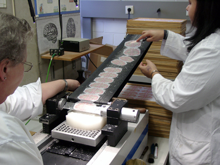
“Constructing BigBrain was a pain-staking process of many years,” says Dr. Alan Evans, a researcher in The Neuro’s McConnell Brain Imaging Centre, a co-founder of the International Consortium for Brain Mapping, and a co-creator of BigBrain. “The atlas is reconstructed from 7,400 brain sections – each as thin as saran wrap. These thousands of sections, many of which were torn or wrinkled, had to be specially stained to reveal their cell bodies, and then carefully re-assembled. Our team developed and employed cutting-edge computational techniques to digitize and compile the brain sections into a 3D atlas.”
Previous histology atlases of the brain were two dimensional and current 3D MRI atlases only have a 1mm resolution. Zooming into an MRI (Magnetic Resonance Imaging, which is a test that uses a magnetic field and pulses of radio wave energy to make pictures of organs and structures inside the body) provides no further detail because the image just reveals the blocks of pixels. BigBrain’s data resolution in each dimension is 50 times more powerful than a regular MRI scan, with an image data set that is 125,000 times bigger. BigBrain’s volume is gargantuan at 1,000 GB or one terabyte.

The 3D Brain atlas is reconstructed from 7,400 brain sections – each as thin as saran wrap.
The research and medical world will have free, easy access to BigBrain’s data, which will be available on the internet at www.bigbrain.loris.ca. It can be used to correlate and integrate widely different types of data: genetic, molecular electrophysiological and pharmacological among many. It will also enable the construction of computer models that simulate various brain functions, normal development and degeneration caused by disease.
In addition, the BigBrain atlas will improve the interpretation of low-resolution in-vivo images obtained from MRI and PET (Positron emission tomography, which is a nuclear medical imaging technique that produces a three-dimensional image or picture of functional processes in the body) scans, by combining the data with the enormous detail and resolution of the atlas. Neurosurgeons will benefit from Big Brain’s 3D information when they, for example, place stimulators deep in the brain during operations for Parkinson’s disease patients. It will also advance clinical research, for example researchers will be able to use BigBrain to locate the site of specific nerve cells that contribute to epilepsy.
“With this atlas, the potential for exploring and analyzing the brain is virtually limitless,” says Dr. Evans. “I think this is only the beginning to unlocking the many secrets of the human brain, which will lead to better research, treatment and ultimately patient care.”
