Be still our beating hearts: Electrophysiology Lab at the Montreal General
 There was some bustle in the E5 wing at the MGH – people were celebrating the opening of the brand new cardiac electrophysiology laboratory (EP lab). “It took a lot of hard work and planning,” said MUHC Director of Cardiac Electrophysiology, Dr. Vidal Essebag, “but the medical and project management teams involved did a wonderful job and we have our lab - the improvement to patient care will be tenfold.”
Cardiac Electrophysiology is the study of the electrical activity of the heart and disorders of heart rhythm. Usually, these studies are performed to diagnose and treat arrhythmias (a heart that beats too quickly or too slowly). There are varying degrees of arrhythmias: some are harmless, but others can be lethal. “We’ll be able to help a lot of people now that we have an EP lab,” explains Dr. Essebag.
Indeed, with 1,000 procedures performed every year, the MUHC will be able to provide better care to its community. “The new lab will improve patient care and shorten wait times. This state of the art EP lab is the first of its kind at the MUHC and will allow us to provide onsite curative procedures for a wide range of complex arrhythmias for the entire population served by McGill affiliated hospitals,” states Dr. Essebag. The new electrophysiology lab, in addition to improving care, will improve teaching and research too. Thanks to the new laboratory, medical residents will be able to witness procedures being performed live, and the MUHC and its medical professionals will have the chance to participate in international studies on the latest therapies.
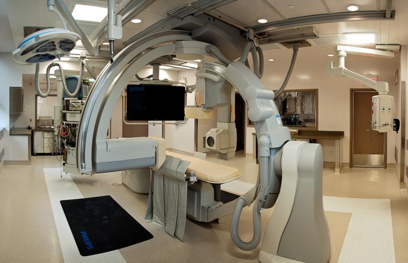
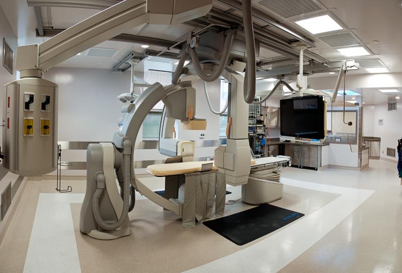
 Nestled at one end of the E Wing, the EP lab is full of cutting-edge equipment; “Our lab is equipped with a built-in bi-plane fluoroscopy machine for x-rays and a three-dimensional mapping system,” explains Dr. Essebag. This means that doctors can see the heart in two dimensions: front to back and side to side. This will allow them to better perform complicated and intricate procedures. “I can see the heart from different angles at the same time and with the three-dimensional mapping system, I can re-create the anatomy of a patient’s heart.”
Essentially, this means that doctors create a map of the heart and determine where an arrhythmia is situated by measuring the amount of electricity the heart generates and the source of abnormal heart rhythms. Once the origin of the irregular heartbeat is found, medical teams can either perform a radio-frequency ablation (burn the extra tissue causing the arrhythmia) or a cryo-ablation (freeze the tissue).
“It’s phenomenal that we have this technology. Having the tools to provide better care is essential to everyone, and now we have them,” says Dr. Essebag.